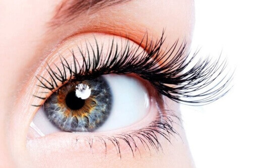
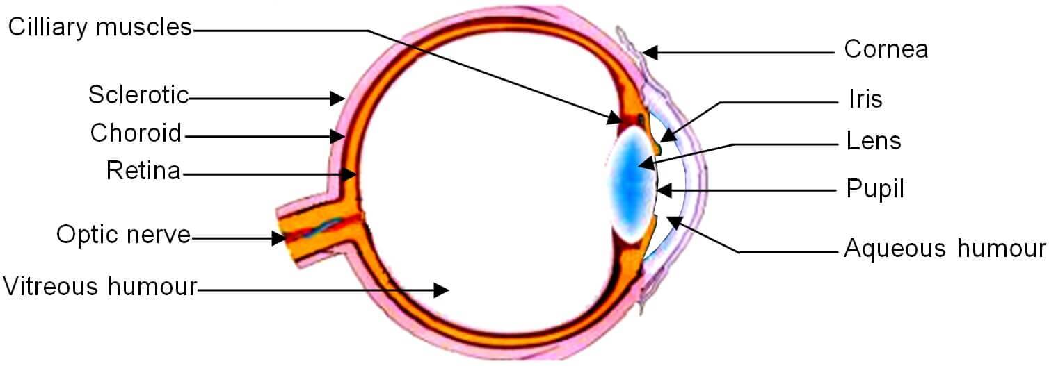
The human eye is one of the most important and sensitive sense organ gifted to us by the God. The construction and working of the human eye is like a camera forming an inverted and real image. The diagram showing the structure of human eye are sclerotic, cornea, choroids, iris, pupil, eye lens, cilliary muscles, retina, aqueous chamber, vitreous chamber, optic nerve, blind spot and yellow spot.

Structure and Working of Human Eye
The human eyes are spherical balls like structures of diameter 2.5 centimeters. These are present in bony orbital cavities. Following is the detail of different parts of a human eye.

1. Sclerotic
The human eye consists of 3 layers. Sclerotic is the outermost layer of human eye. Its function is to protect the eye from the mechanical injuries. A part of sclera is visible in front of the eye as white.
2. Cornea
Cornea is the transparent part of the sclerotic. It is slightly bulged out in front of the eyeball. The function of cornea is to admit light into the eye.
3. Choroid
Choroid is the second layer of eye which is present between sclera and retina. It is opaque and tough.
4. Iris
Iris is an opaque, dark and muscular diaphragm which is present behind the cornea. There is a hole in the middle of the iris which is called ‘pupil’. The pupil appears black and its size is controlled by the iris. Thus the main function of iris in the eye is to regulate the amount of light entering the eye through pupil by controlling the size of pupil. If the light is bright then the pupil contracts so that less light can enter the eye. On the other hand, if the light is dim then the pupil expands so that more light can enter the eye.
5. Eye lens
The eye lens is an elastic, transparent and biconvex structure. It is present behind the iris. It is held in its position by means of cilliary muscles. The focal length of the eye lens can be changed by stretching or relaxation of cilliary muscles. Thus cilliary muscles help the eye in changing the focal length of eye lens so that the eye can observe both the nearby and distant objects. The ability of the eye to see the distant objects as well as the nearby objects by changing the focal length of eye lens is called accommodation.
6. Aqueous chamber
The space between the cornea and eye lens is called aqueous chamber. It is filled with a viscous and watery fluid called aqueous humour.
7. Vitreous chamber
The space between eye lens and retina is called vitreous chamber. It is filled with a transparent, gelatinous material called vitreous humour.
8. Retina
Retina is third and innermost light sensitive layer of eye on which the image is formed. Retina contains a large number of light sensitive cells called ‘rod cells’ and ‘cone cells’. The rod cells respond to the intensity of light, while the cone cells respond to the colours present in the light. The cone cells present in the retina are of 3 types:
1. Red sensitive
2. Blue sensitive and
3. Green sensitive.
Actually when an image formed on retina, the rod cells and the cone cells get stimulated and send the information in the form of electrical signals to the brain through the optic nerve and give rise to the sensation of vision. The area of retina where image is formed is called yellow spot. On the other hand, the area of retina over the exit of optic nerve does not contain rod cells and cone cells, so it does not responds to the light so it is called blind spot.
Test Your Understanding and Answer These Questions:
- Define yellow spot.
- Define blind spot.
- What is retina? What is its function?
- What is the function of sclerotic in human eye?
- What is the function of cilliary muscles in human eye?
- What type of lens is present in human eye? What is its main function?
- Explain the structure of human eye with the help of a well labeled diagram.
- What are rod cells and cone cells? What is function of rod cells and cone cells?
