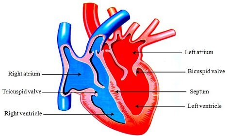
The human heart is a conical, hollow and muscular organ which helps in pumping blood around all the parts of body. It is covered by a two layered membrane called pericardium. The function of pericardium is to protect heart from shock and mechanical injuries.
Internally, the human heart is divided into four chambers. The upper two chambers of heart are called atria (singular atrium) or auricles,. The lower two chambers of heart are called ventricles. The atria (or auricles) are further classified as left atrium (left auricle) and right atrium (right auricle). In the same way, ventricles are also classified as left ventricles and right ventricles.
Atria open into ventricles of their sides. The openings between atria and ventricles are guarded by valves. The valve present between left atria and left ventricle contains two flaps and is called bicuspid valve. The valve present between right atria and right ventricles contains three flaps and is known as tricuspid valve. The function of these valves is to prevent the backflow of blood from ventricles into atria when the ventricles contract. The right atrium and ventricle are separated from left atrium and ventricle by a partition called septum.

Working of Heart
The function of heart is to pump the blood around all the parts of body. This is done by regular contraction and relaxation of the atria and ventricles. The contraction and relaxation of the atria and ventricles take place alternately. One complete contraction and relaxation of the heart is called heart beat. The heart of a normal healthy person beats about 70 to 72 times per minute under normal conditions. But, the rate of heart beat can increase during special circumstances. For example, during hard physic exercise the rate of heart beat can be more than 100 per minute.
Now, we shall discuss how the heart circulates blood in our body.
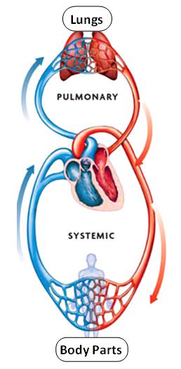
 1. The muscles of right atrium relax and it receives deoxygenated blood from all the parts of the body. The deoxygenated blood is forced into right ventricle, by the contraction of right atrium.
2. When the right ventricle contracts, the de-oxygenated blood is forced into lungs. During the contraction of right ventricle the backward flow of blood into right atrium is prevented by the bicuspid valve. In the lungs, the deoxygenated gives up carbon dioxide and picks up oxygen. So, in lungs the blood becomes oxygenated.
3. Then the muscles of left atrium relax and it receives oxygenated blood from the lungs. The oxygenated blood is then forced into left ventricle, by the contraction of left atrium.
4. When the left ventricle contracts, the oxygenated blood is pumped to all the parts of the body except lungs. During the contraction of left ventricle the backward flow of blood into left atrium is prevented by the tricuspid valve. In the body, the oxygenated blood gives up oxygen to the cells and takes back carbon dioxide from them. So, in the body the blood becomes deoxygenated.This deoxygenated blood then flows back to the heart and enters by the right atrium. In this way, the whole process is repeated again to ensure continuous flow of blood in the body.
Test Your Understanding and Answer These Questions:
- What is heart beat?
- Explain working of human heart.
- Explain structure of human heart.
- What is the function of valves in heart?
- What is the affect of exercise on heart beat?
- Draw human heart and label its various parts.
- Name the membrane that covers human heart
